
Result 1: MSNs markers are detectable by DAB Staining
By IHC I showed that both antibodies work and both markers are expressed in many striatal neurons whereas they are not expressed in cortical neurons.
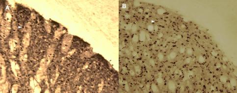
Expression of the MSNs markers in the striatum of an mGluR5KD-D1 mouse. A) Detection of DARP-32 B) Labling pre-pro Enkephalin (ppENK)
Result 2: The mGluR5KD-D1 is only expressed in D1-R expressing neurons
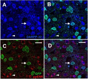
IFC labeling showing that the expression of the transgene is selective to D1-MSNs
A) DAPRP-32 was detected in all D1R and D2R (blue).
B) Green Fluorescent Protein (GFP) which is introduced into the transgenic mice under D1R promoter was only detected in around half of the MSNs (green).
C) Pre-pro Enkephalin (ppENK) which is only expressed in D2R was labelled in almost half of the MSNs (red) and by merging the photos no green and red cell bodies were colocalozed.
D) Merging all sections shows that all the blue cells express either red or green markers but no colocalization of red and green is found.
Responsible for this page:
Director of undergraduate studies Biology
Last updated:
05/24/10