
The transgenic mouse model
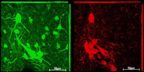
After genotyping the mice using PCR, double immunolabelling with anti-ChAT (choline-acetyltransferase) and anti-GFP was performed. If the expression system in this model is stable, this means that the staining should overlap in all cholinergic neurons, including the audio-vestibular efferent neurons. I found an overlap in the vestibular efferents (see figure), but not in either of the two types of olivocochlear efferents (MOC or LOC neurons) (data not shown). Further, I found strong double labelling of motor neurons of the trigeminal nerve (5th cranial nerve), the abducens nerve (6th cranial nerve) and the facial nerve (7th cranial nerve) (data not shown).
The lack of eGFP expression in the olivocochlear neurons indicates that this mouse model is unsuitable for targeting those efferent neurons. However, the vestibular efferents express eGFP and the model can therefore be used for targeting these efferents when performing electrophysiological experiments on living cells. During such experiments the neurons can be identified by eye in a fluorescence microscope and subsequently recorded from. Another use for the mouse model would be for targeting of motor neurons, which show robust eGFP expression in all motor neuron nuclei existing in our tissue.
Kv4.3 expression
The Kv4 family of potassium channel subunits consists of Kv4.1-3. I found Kv4.3 to be expressed in vestibular efferents and in both types of olivocochlear efferents, as well as in principal cells in the LSO (see figure). Kv4.2 was not found above background level in any of the neurons of interest and Kv4.1 has previously been shown not to be expressed in the areas of interest (Serodio and Rudy, 1998; Fitzakerley et al., 2000). This leads to the conclusion that the the channels causing transient outward currents observed in olivocochlear efferents are built up from Kv4.3 subunits. Combined with the results from preliminary electrophysiological experiments (Anna K MAgnusson, unpublished data) it can be concluded that the vestibular efferents probably also have these transient outward currents and that they are also in these neurons built up of Kv4.3 subunits.
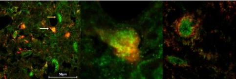
Since a pre- or post-synaptic localization of the potassium channels can be of biological relevance for the cell, I investigated this by double staining against Kv4.3 and the pre-synaptic neuronal marker SV2A. I found that there is no overlap between the Kv4.3 and the SV2A, meaning that Kv4.3 is expressed post-synaptically in lateral olivocochlear efferents and principal cells of the LSO (see figure).
Responsible for this page:
Director of undergraduate studies Biology
Last updated:
05/18/10