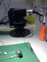
Tissue Viability Imaging (TiVi)
Tissue viability Imaging gives information about the skin microcirculation by using subsurface polarization light spectroscopy (Henricson et al. 2009). The technique utilizes a digital camera (EOS 55D, Canon) equipped with perpendicular polarization filters. When light flashes from the camera, white light gets polarized by the polarization filter. The reflected light from the skin contains both the same polarized light and randomly polarized light. When the light falls on the skin, a part of the light gets reflected directly by the superficial layers of the skin. The major part of the light gets randomly polarized due to the backscattering of other dermal tissues and part of the light is absorbed. Directly reflected light from the skin is filtered out using a second polarizing filter on the camera lens. Red Blood Cells in the skin absorb light in the 500 – 600nm spectral range (green light region) whereas the other tissues absorb lights of other wavelengths. Hence the TiVi method can distinguish the images according to their wavelength absorption ranges. Being a standard photographic technique, TiVi is not sensitive to motion and provides an instantaneous picture.
Experimental Protocol
TiVi tests were conducted with 7 Red Jungle fowls and 4 Zebra finches (non-broody birds). Initially, Jungle fowls were anesthetized with 4% Isoflurane and 3% for Zebra finch. A specific area on the brood patch was marked with a permanent pen to make sure that the pictures were taken on the same location. As a control, three photographs were taken at the interval of 10 seconds by applying Klick gel. Then the NO gel was applied on the marked area using cotton bud and left to give an effect for one minute. The gel was gently wiped out using soft tissue and repeated pictures were taken again with an interval of 10 seconds. The data was analyzed using “Wheels bridge” software.
Responsible for this page:
Director of undergraduate studies Biology
Last updated:
05/19/12
