
Introduction
Visual acuity is commonly defined as the ability of the eye to resolve the smallest distance between two objects or the ability to resolve spatial details (Land & Nilsson, 2002). Which degree of visual acuity is dependent on different factors but mainly on how many photoreceptors that are connected to a single ganglion cell (Land & Nilsson, 2002). The photoreceptor/ganglion cell ratio varies across the retina (Land & Nilsson, 2002). Increasing distance between ganglion cells in the retina indicates a higher signal summation from several photoreceptors and high convergence, therefore lowers the ability to achieve high visual acuity. In cats the highest ganglion cell density and thereby the highest visual acuity is obtained in the central region of the retina, called the area centralis (Wässle & Boycott, 1991). The retinal ganglion cells can also be distributed in a dense elongated arrangement, a horizontal visual streak (Peichl, 1992). Such an arrangement is found in e.g. wolves (Peichl, 1992).
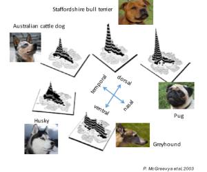
Interestingly, Mcgreevy and colleagues (2003) showed that the skull shape of dogs correlate with ganglion cell topography. Dolichocephalic breeds (with long skulls) have a horizontal visual streak with high visual acuity, while brachycephalic breeds (with short skulls) have ganglion cells more concentrated in a round area centralis (Mcgreevy et al, 2003). The area centralis is shown to have approximately three times more ganglion cells (2640/mm2) than in the horizontal streak (880/ mm2), which gives the brachycephalic dogs an advantage in visual acuity over dolichocephalic breeds (Mcgreevy et al, 2003). However, to our knowledge, it has not been investigated whether the differences in ganglion cell distribution correlate with the perceived visual acuity in dogs of different skull shapes. Additionally, very few studies have investigated the visual acuity of dogs in dim conditions.
The retina of the dog consists in majority of rod photoreceptors operating in dim light conditions. Even though there is a higher number of cones in the more central parts of the retina, the majority of photoreceptors here is rods (Mowat et.al. 2008). This distribution is similar to their ancient relative the wolf (Peichl, 1992). The distribution of photoreceptors among breeds and dogs of different skull shape are not yet known. If we assume that the photoreceptor distribution is the same in both brachycephalic and dolichocephalic dogs then, since brachycephalic dogs have higher number of ganglion cells in the area centralis, they schould outperform the dolichocephalic breeds in visual acuity also in dim conditions.
The aim of this study is to determine possible visual acuity differences in breeds with extreme skull shapes, in both daylight and dim light conditions. The best way to assess what an animal really perceives is to perform behavioural tests. Therefore, this study will use two representative breeds in a two-alternative discrimination test, which has shown to be successful when to determine sensory abilities in animals (Roth et.al. 2008). The test will be performed in daylight to test the visual acuity generated by the cones and in dim light to test the visual acuity generated by the rods. I expect the dolichocephalic breed to perceive lower visual acuity than the brachycephalic breed at both light intensities due to that their horizontal visual streak have less ganglion cells to sum up the visual signal and therefore lowers their visual acuity.
Responsible for this page:
Director of undergraduate studies Biology
Last updated:
05/03/16