

Beta cell plasma membrane lipid separation bands from HPTLC plates are shown after Orcinol (left) and Primulin spray (right).
Here, we are trying to re-establish the unpublished work done by Professor Spitalnik, Columbia University in 1991 that are shown in the below.
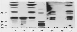
RINm5F total lipid extract. Lane 2 & 6 RINm5F Folch upper phase’s lipids. Lane 3&7 RINm5F Folch lower phase’s lipid. Lane 4 human Brain glycolipid. (Fig- Spitalnik et al, 1991). But in his work there were some missing experiments that are essential to prove our hypothesis. Professor Spitalnik has done his experiments on RIN 5AH cell lipids. As sulfatides are found on the plasma membrane, it is good idea to work with plasma membrane lipids. So, we have done our experiments on both from plasma membrane lipids and cell lipids.To get better comparable result, we have done our experiments with different cell lines besides pancreatic beta cell lines. To re-establish the hypothesis that IC2 binds with the auto antigen on the pancreatic beta-cell plasma membrane, we are just far from the last immunobloting step
Responsible for this page:
Director of undergraduate studies Biology
Last updated:
05/20/09