
Hormone analysis
Corticosterone

CORT analysis was conducted using ELISA kits from ENZO Life Sciences using 96 well plates. Blood plasma samples were thawed at room temperature for 30 minutes prior to beginning the procedure and the ELISA kit was warmed at room temperature for the same period of time. Prior to starting the experiment assay buffer, wash buffer and 1:100 steroid displacement reagent (SDR) was prepared. Blood plasma samples were diluted to requirement and standards of known CORT concentration (pg/mL) (20000, 4000, 800, 160, 32) were prepared immediately before the experiment and used within 1 hour of mixing. 150 µl assay buffer was added to non-specific binding (NSB) wells. 100 µl assay buffer added to Blank wells. Standards and Samples were added to assigned wells. Conjugate was added to all wells except Total and Blank wells. Antibody was then added to all wells except Total, Blank and NSB wells. The well plate was then covered with a plastic sheet and placed on plate shaker for 2 hours incubation at 500 rpm at room temperature. The plate was then aspirated and washed with wash buffer adding 200 µl wash buffer to each well 3 times, inverting between each time and patting dry. 5 µl of Conjugate was then added to Total wells. 200 µl pNpp Substrate added to all wells, covered with plastic and aluminium foil to block the light. Well plate incubated for 1 hour without shaking at room temperature. Stop solution was then added all wells to stop reaction and absorbance at 405 nm immediately read using a plate reader. Samples were then compared to a standard curve; CORT concentrations were extrapolated and then multiplied by their dilution factor.
Testosterone and estradiol
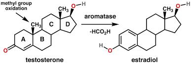
Testosterone and Estradiol were analysed using corresponding ELISA kits from MyBioSource using the same protocol for both hormones. Samples were thawed and all reagents were warmed to room temperature before use for a period of 30 minutes. A wash buffer was prepared by diluting 15 ml of concentrated wash buffer with 285 ml deionised water. Firstly the Blank wells were assigned to the plate, 50 µl of Standard or Sample were then added to their assigned wells. 50 µl HPR-Conjugate was added to all wells except Blank and then 50 µl Antibody was added to all wells. The plate was then covered with adhesive film and incubated for 1 hour at 37 °C. All wells were then aspirated three times with 200 µl wash buffer administered using a multichannel pipette. Liquid was completely removed each time by inverting plate gently and patting dry. 50 µl Substrate A and 50 µl Substrate B was added to all wells, mixed well; then incubated for 15 minutes at 37 °C. The plate was covered in aluminium foil for this incubation period in order to block light. Finally 50 µl of Stop Solution was added to each well, the highest concentrated standards should develop an obvious blue colour. Optical density was then determined by using a microplate reader set to 450 nm and hormone levels were compared to the standard curve.
Responsible for this page:
Director of undergraduate studies Biology
Last updated:
05/20/18